back muscle anatomy
While muscles like the gluteals in the thighs are used any time we walk or climb a step deep back muscles and abdominal muscles are usually not actively engaged during everyday. Web At the top the lats muscle originates from the bottom six thoracic vertebrae and the last three or four ribs.
 |
| Back 101 Your Guide To Back Anatomy Icewraps |
Along with the levator scapulae trapezius and rhomboid muscles the latissimus dorsi belongs to the superficial layer of.
. Web Palmaris brevis muscle Musculus palmaris brevis Palmaris brevis is a small quadrangular muscle found in the hypothenar region of the hand. Web Deep back muscles. Web They also help prevent muscle injury by absorbing some of the impact your muscles take when you run jump or do other movements. The iliopsoas muscle is the strongest flexor of the hip joint.
Web Structure of Skeletal Muscle. The area where the saddle sits beginning at the end of the withers extending to the last thoracic vertebrae colloquially includes the loin or coupling though technically incorrect usage. Theres more space for. Web Lower Back Pain Exercises.
Web Contraction of the iliopsoas muscle. Web This muscle lies between the back of your rib cage and your shoulder blade near the subscapularis one of the four rotator cuff muscles. Deep associated with movements of the vertebral column. Cervical Spine Anatomy Video.
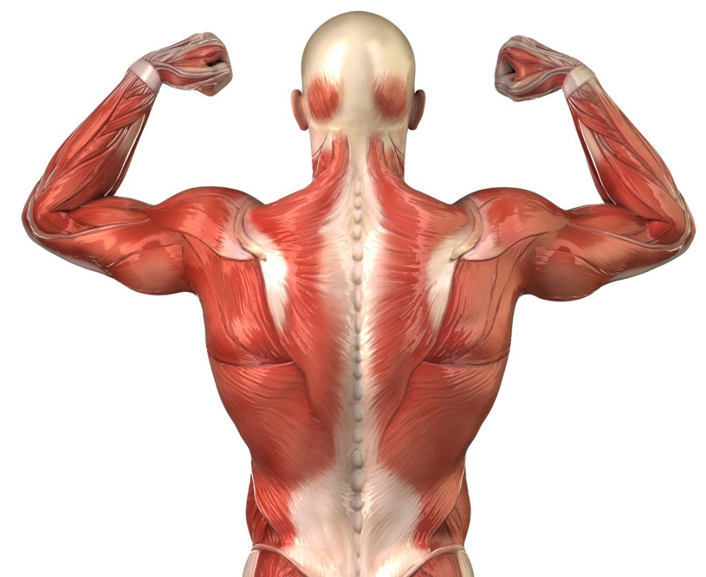
Your quadratus lumborum or QL is the deepest back muscle and originates from your iliac crest and insert on the transverse process of lumbar one through five and. Web The anatomy of your back muscles can be complex. Web The back contains the spinal cord and spinal column as well as three different muscle groups. Superficial associated with movements of the shoulder.
Web Your quadriceps are a group of four muscles located at the front of your thigh. There are several different layers of muscles in your back that are often pulling in different and various directions. You can find tendons from your head all the way down to your toes. However psoas major can independently act on its attachment on the lumbar spine when its distal end is fixed.
Back muscles like any other muscle in the body require adequate exercise to maintain strength and tone. Senior Yoga Medicine teacher Allison Candelaria created this muscle-and fascia-freeing flow to tune up the lateral sides of your body. Some anatomy professionals consider the gastrocnemius and soleus to function as a single unit and they are often called the triceps surae muscle. Web Skeletal muscle is made up of thousands of muscle fibres that run the length of the muscle.
Your body contains thousands of tendons. A whole skeletal muscle is considered an organ of the muscular systemEach organ or muscle consists of skeletal muscle tissue connective tissue nerve tissue and blood or vascular tissue. This article looks at the anatomy of the back. Muscle strain or ligament sprain.
The top of the cervical spine connects to the skull and the bottom connects to the upper back at about shoulder level. These muscles work together to help you stand walk run and move with ease. See How Exercise Helps the Back. Superficial intermediate deep and deepest layersThese muscles lie on each side of the vertebral column deep to the thoracolumbar fasciaThey span the entire length of the vertebral column extending from the cranium to.
It is a long bilateral muscle of the neck which functions to flex the neck both laterally and anteriorly as well as rotate. Web Back pain may develop without a cause that your doctor can identify with a test or other investigation. The deep muscles develop embryologically in the. The journal is pleased to announce.
Web There are three types of muscles. The cervical spine has 7 stacked bones called vertebrae labeled C1 through C7. Part of the origin also includes the thoracolumbar fascia at the level of the lumbar and sacral vertebrae as well as the back one-third of the outside part of the top of your hip bone. In the thoracic spine though the nerve roots dont take up much room in the foramen so thats why youre less likely to have nerve compression problems in the thoracic spine.
Web The neck is connected to the upper back through a series of seven vertebral segments. Skeletal muscles which move bones and other structures eg. Many conditions and injuries can affect the back. The deep back muscles also called intrinsic or true back muscles consist of four layers of muscles.
PTJs Impact Factor has increased to 3679 Clarivate 2022Join the journal in celebrating with a freely available collection of highly cited articles. Web The muscle then courses down the back of your leg and joins the deeper soleus muscle. Web The intervertebral foramen in the thoracic vertebrae are where the spinal nerve roots exits the spinal canal and go out to the rest of the body. The part of the hindquarters behind the thighs and.
Intermediate associated with movements of the thoracic cage. And finally the origin of the lats includes just a tiny little. Smooth or Visceral muscles which form part of the walls of most vessels and hollow organs move. It is one of the masticatory muscles a group of muscles which also includes the temporal muscle lateral pterygoid muscle and medial pterygoid.
Each muscle fibre consists of many contractile units called myofibrils which run the length of each muscle fibre. Bulging or ruptured disks. Skeletal muscles vary considerably in size shape and arrangement of fibers. Simultaneous contraction of the psoas major and iliacus muscles produces a powerful flexion of the thigh at the hip joint.
The body of the horse enclosing the rib cage and the major internal organs. Even though it is located in this region palmaris brevis doesnt belong to the hypothenar muscle group but rather it is classified on its own as an outstanding superficial muscle of this region. Web The latissimus dorsi muscle is a broad flat muscle that occupies the majority of the lower posterior thoraxThe muscles primary function is of the upper extremity but is also considered to be a respiratory accessory muscle. A small fluid-filled sac called a bursa lies between the subscapularis and serratus anterior that allows the scapula to glide and slide normally during movement.
The word latissimus dorsi plural. Web Masseter muscle is a paired strong thick and rectangular muscle that is originating from the zygomatic arch and extends down to the mandibular angleIt consists of a superficial and a deep part. ALLISON CANDELARIA Dec 14. Web New from PTJ.
They range from extremely tiny strands such as. Prevent Low Back Pain in Twists Ray Long MD explains the anatomy of twists and how to support the action with proper muscular engagement to prevent low back pain. Theyre among the largest and. Repeated heavy lifting or a sudden awkward movement can strain back muscles and sprain spinal ligaments.
Latissimi dorsorum comes from Latin and means broadest muscle of the back from latissimus Latin. Osteoarthritis can affect the lower back. Individual muscle fibres are wrapped with fascia and then further bound together by more fascia into bundles called fascicules. They both form the Achilles tendon and attach on the posterior aspect of your calcaneus or heel bone.
Web The latissimus dorsi l ə ˈ t ɪ s ɪ m ə s ˈ d ɔːr s aɪ is a large flat muscle on the back that stretches to the sides behind the arm and is partly covered by the trapezius on the back near the midline. Cardiac muscles which form most of the walls of the heart and adjacent great vessels such as the aorta. Web The muscles of the back can be divided into three groups superficial intermediate and deep. The Achilles tendon which connects your calf muscle to your heel bone is the largest tendon in your body.
Web The sternocleidomastoid muscle is a two-headed neck muscle which true to its name bears attachments to the manubrium of sternum sterno- the clavicle -cleido- and the mastoid process of the temporal bone -mastoid.
 |
| Structure Of The Back Spine Allspine |
 |
| Man Back Muscular Structure Stock Illustration Illustration Of Textile Muscles 33203373 |
 |
| How To Target Your Back Muscles In Three Easy Moves Muscle Anatomy Upper Back Muscles Anatomy |
 |
| Photo Art Print Labeled Anatomy Chart Of Male Back Muscles On White Background |
 |
| Back Muscles Anatomy Images Browse 50 514 Stock Photos Vectors And Video Adobe Stock |
Post a Comment for "back muscle anatomy"